Introduction to Optical Imaging
In recent years, brain imaging technology has become a new favorite in the research of cognitive neuroscience, and cognitive neuroscience has made important contributions to the vigorous development of psychology. Several brain imaging techniques have been cited, such as functional magnetic resonance imaging, positron emission computed tomography, single photon emission computed tomography, and optical imaging.
The development of optical imaging technology provides a new important research method for the study of the brain mechanism of cognitive activity. Optical imaging technology can reveal the structure and function information of the nervous system at different levels, and provide an important experimental basis for explaining cognitive activity from a new perspective. The basis of optical imaging is that the activity of neurons will cause the changes of some related substances (such as water and ions), resulting in changes in their optical properties, which are obtained after interacting with the added light quantum of certain specific wavelengths Corresponding optical signal. The imaging instrument system detects the spatial distribution of this optical signal within a certain time interval to form an image. Optical imaging has higher spatial and temporal resolution than fMRI, and can measure changes in total deoxygenated hemoglobin, total hemoglobin, and blood volume with smaller voxels. There are many types of optical imaging, including near infrared spectroscopy (NIRS) and optical coherence tomography (OCT), which can provide high-resolution images for observing functional columns of the cerebral cortex. NIRS can pass through the skull and has been used for noninvasive brain function studies in animals and children.
Application significance of fNIRI
The functional near-infrared optical monitoring technology is a very useful supplement to the existing fMRI and PET technology. It uses the blood volume and blood oxygen in the tissue as the information carrier, by measuring the distribution and changes of blood volume and blood oxygen in the cerebral cortex Situation to understand brain activity. The functional near-infrared optical monitoring technology can perform real-time non-invasive measurement, and has the advantages of high time accuracy, flexibility, ease of use, and low cost. Compared with fMRI, although its spatial resolution does not reach the level of fMRI, it is price, portability, ease of use and almost no interference with the subject (due to the measurement method, fMRI is easy to cause the subject ’s claustrophobia ", Which is not conducive to the detection of brain activity) and other aspects are far better than fMRI. Compared with PET, the functional near-infrared optical monitoring technology does not require additional equipment such as the cyclotron necessary for PET and the operation of injecting nuclear radiation into the human body, which is more beneficial to the human body in terms of safety and can be carried out on the subjects. Multiple long-term measurements. In addition, the fMRI and PET technologies require that the subjects cannot move during the experiment, so it is difficult to study children. The functional NIR optical monitoring technology does not have this limitation. These make the functional near-infrared optical monitoring technology have great advantages in the study of large-scale brain activity, which has aroused widespread concern in the international neurobiological community. fNIRI system composition and working principle
Near-infrared brain functional imaging technology (fNIRI) can provide cerebral oxygenation information of cerebral cortex during brain functional activity-oxygenated hemoglobin concentration change (△ [oxy-Hb]), deoxyhemoglobin concentration change (△ [deoxy-Hb]) Hexue (△ [tot-Hb]) is a new technology developed in recent years. It has the characteristics of low price, portability, and real-time non-invasive measurement. It has been more and more widely used in brain research and clinical testing. Praise. In China, the Key Laboratory of Biomedical Photonics of the Ministry of Education of Huazhong University of Science and Technology has developed a portable near-infrared brain functional imager based on continuous light, which can sensitively detect the blood changes of the prefrontal lobe activation in the advanced functional area of ​​the brain. Used in language research to achieve some meaningful results.
Near-infrared spectroscopy is an important content in the field of biomedical photonics. It is based on the absorption spectra of major chromophores (oxygenated and reduced hemoglobin, cytochrome oxidase, etc.) in living tissues, combining light in tissues. Propagation law, using the near infrared light to penetrate the tissue, to study the tissue biochemical information related to the absorption spectrum carried by the light after a series of absorption and scattering in the tissue, the main purpose is to study these absorption in the tissue The quantitative measurement method of chromophore concentration is the basis of research on tissue hemodynamics, tissue optical monitoring, and functional monitoring. When a certain area of ​​the cerebral cortex is active, its local blood volume will increase, and the increase reflects the degree of activation. Therefore, the amount of blood volume change in the cortex can be used as an indicator of brain functional activity. The brain function monitoring system based on near infrared spectroscopy can non-invasively measure the concentration changes of oxyhemoglobin and reduced hemoglobin in a certain area in the cerebral cortex in real time, so that the changes in blood oxygen and blood volume in this area can be calculated, Monitor the functional activity of the cerebral cortex.
Oxygen is the basis of all life activities. The same is true for human brain tissue, which is accompanied by complex oxygen metabolism processes. Real-time monitoring of the blood dissolved oxygen concentration in brain tissue can realize the peeping of brain activity functions to obtain brain activity Real information. Studies have shown that during brain activation, the level of excitability of neural activity increases, and local cerebral tissue blood flow, blood volume, and blood oxygen consumption all increase, but the increase rate is different, and the increase in cerebral blood flow exceeds the blood flow volume 2 ~ 4 times, while the oxygen consumption increased only slightly, and the blood flow increased beyond the increase in oxygen consumption. This difference led to an increase in the venous blood oxygen concentration in the brain activation function area, and a relatively reduced deoxyhemoglobin. Using near infrared spectroscopy ( NIRS) enables real-time, non-invasive, in-vivo measurement of parameters such as the relative concentration changes of major chromophores in superficial tissues such as oxyhemoglobin (HbO2), deoxyhemoglobin (Hb), cytochrome oxidase (CytOx), and blood concentration , Through a certain image restoration and reconstruction can further obtain near-infrared optical images of brain activity. Near-infrared optical imaging (fNIRI) is a new type of brain functional imaging technology that appeared in the 1990s fNIRI uses near-infrared spectroscopy to record changes in blood oxygen and blood volume parameters at different locations in the brain to obtain brain function images.
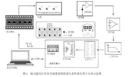
The composition and principle of the fNIRI system are shown in Figure 1. It is mainly composed of three parts: a flexible probe, a measurement and control module and a computer.

The control module includes a light source constant current drive unit, a post-amplification filter unit and a data acquisition part. Under the control of the clock frequency set by the computer, the light source drive unit adopts time division multiplexing technology to light up the four light sources on the probe in sequence. The light source emits The near-infrared light of a specific wavelength illuminates the tissue to be measured, and then the detector is responsible for receiving the optical signal after tissue attenuation, and completing the photoelectric conversion and signal pre-amplification. The front-end analog signal is further amplified and filtered and processed in data acquisition The analog-to-digital conversion is completed in the card, and finally input into the computer through the USB interface. The computer uses the system software to control the working state of the circuit, and performs real-time calculation on the collected multi-channel data, and displays the bleeding oxygen / blood on the interface The volume concentration changes to reflect the functional activity of the brain regions corresponding to each channel.
Advantages and disadvantages of fNIRI
The advantages of the fNIRI system are flexibility, ease of use, low cost, real-time, and non-invasiveness. Although the spatial resolution is not as good as functional magnetic resonance imaging (fMRI) and positron emission tomography (PET), etc., it is characterized by its ability to recognize Real-time functional imaging in active natural scenarios, and can be measured simultaneously with other brain function research methods such as fMRI, PET, EEG, etc. In addition, it is easy to repeatedly experiment on a large number of subjects. FNIRI has become the 1990s One of the most popular research topics in the international neurobiology community. Luo and Chance and others have successfully developed a brain functional optical imager based on near-infrared spectroscopy in the world, and obtained fMRI in the motor area of ​​normal human cortex. Consistent experimental results. Science and Trends in Neuroscience have reported and commented on relevant results.
Compared with the traditional functional magnetic resonance imaging, positron emission tomography, brain electromagnetic detection and other technologies, this technology can not only provide completely nondestructive brain function detection, but also has a very high temporal resolution and ideal spatial resolution. The near-infrared spectroscopy system has the advantages of small size, light weight, portability, and low cost. It has great application value in the field of cognitive neuroscience researchers' wide attention to brain function research.
Examples of application of fNIRI in psychology
At present, NIR spectroscopy has been widely used in the monitoring of brain functional activities. With the advancement of photoelectric technology and information technology, near infrared optical monitoring has been further developed. In this field, breakthroughs have been made in the study of infant brains, because infant brains are small in size, simple in geometry, and have regular optical parameters. Lsobed et al. Used near infrared spectroscopy to study the functional activity of neonatal sensorimotor cortex. In the field of cognitive neuroscience, near-infrared brain function monitoring technology is also widely used. Sato et al. Used near infrared spectroscopy to study cortical activity during speech recognition.
As mentioned earlier, the Key Laboratory of Biomedical Photonics of the Ministry of Education of Huazhong University of Science and Technology has developed a portable near-infrared brain function imager based on continuous light, and launched research on various brain functions. For example, in order to detect the activity of the prefrontal lobe of the brain, they carried out a working memory study based on the n-back operation paradigm. The information execution control process of the working memory central execution system is relatively complicated, and its specific content involves different cognitive components such as attention and suppression, task management, planning, supervision management, and information coding. It is difficult to strictly distinguish these different functions in the imaging study of brain function.The study uses the verbal n-back operation paradigm.The main purpose is to use the fNIRI system to test the prefrontal lobe of the subjects when performing the verbal n-back task. The cortical activation status was monitored, and the brain activation data and behavior performance data of the subjects were analyzed. A practical three-wavelength near-infrared brain functional optical imaging system was developed, and its working principle was introduced. A speech-based n- Back working paradigm of working memory research, monitoring the activation of the prefrontal lobe (PFC), analyzing the behavioral performance data (response time and correct rate) of the subjects, and examining the behavioral performance of the subjects and the working memory load effect of PFC-activated brain regions The relationship between the PFC activation amount and behavioral parameters of the subjects under high memory load was discussed. The results show that by measuring the changes in blood oxygen parameters and using near-infrared optical imaging methods, the activity of the cerebral cortex can be monitored in real time.
Another study concerns developmental dyslexia. Developmental dyslexia refers to a phenomenon of learning disabilities in which children have normal intelligence and equal educational opportunities, but their reading scores are significantly behind their age. According to the different results obtained by monitoring the brain function of dyslexic patients with other functional monitoring technologies, an appropriate experimental paradigm was designed to study the cerebral cortex activity of Chinese children with dyslexia during the processing of Chinese character speech and semantics. Abnormal activity of the left prefrontal cerebral cortex in patients with dyslexia provides experimental evidence for neurophysiological studies of dyslexia.
Dog Chew Toys
SAFE DOG CHEW TOYS: Our dog toys are made of natural and non-toxic material that ensures the safety of dogs. Thicker fabric and better stitching make these toys more durable for dogs. It's suitable for teeth cleaning and chewing.
Easy for dogs to carry, toss, and roll around with. Each toy has squeaker inside,it will catch dogs' attention. Your pup will love their new squeaky toys.
HELPS REDUCE ANXIETY IN DOGS and SAVE YOUR FURNITURE:This toys provide your dogs hours of fun.Dogs can chew these toys for hours,dogs will not pay attention to your shoes,cloes and other furnitures.
We are a Chinese pet products manufacturer, our company produces and sells various cat accessories and dog accessories, including Pet Toys , Pet Apparel And Accessories, Pet Collar And Leashes , Pet Beds And Accessories, Pet Cleaning And Grooming Products, Pet Bowls And Feeders , Pet Houses , and Pet Carriers And Travel Products, which are accepted OEM&ODM. Please don`t hesitate to contact me if you need anything.
Dog Chew Toys,Puppy Teething Toys,Tough Chewer Dog Toys,Best Chew Bones For Dogs
Jilin LOYO Pet Products Co.,Ltd. , https://www.loyopetaccessories.com